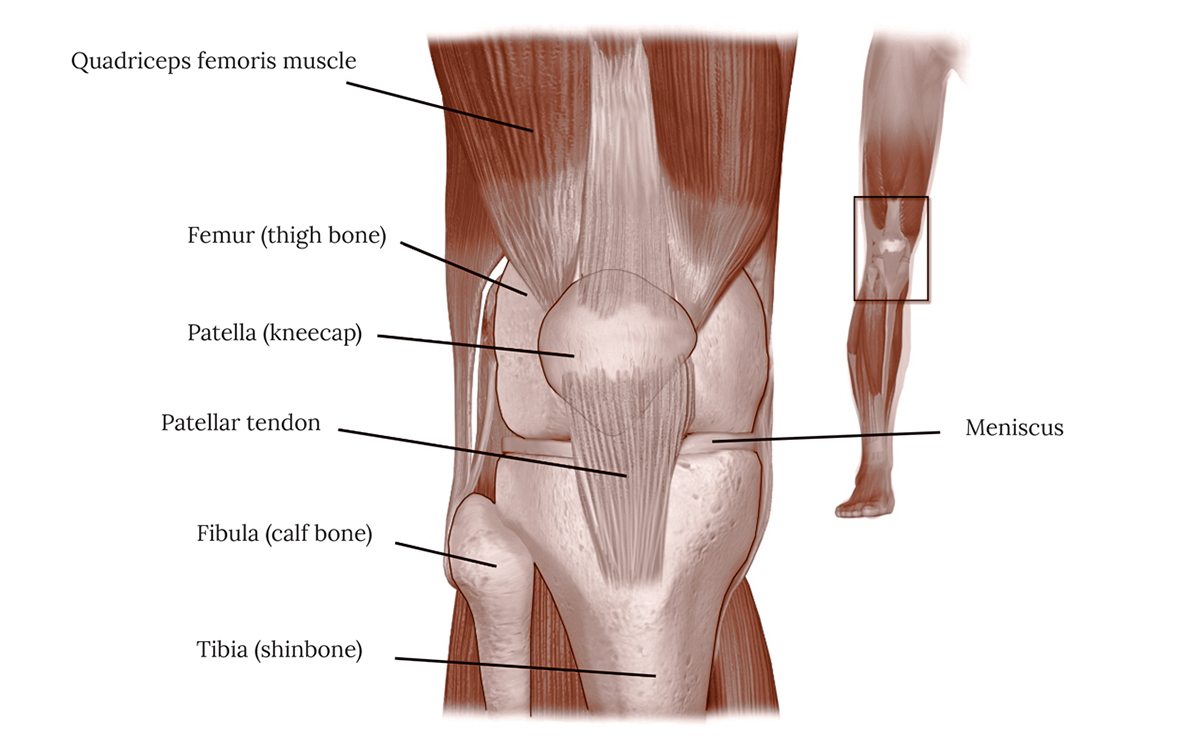
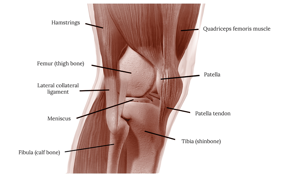
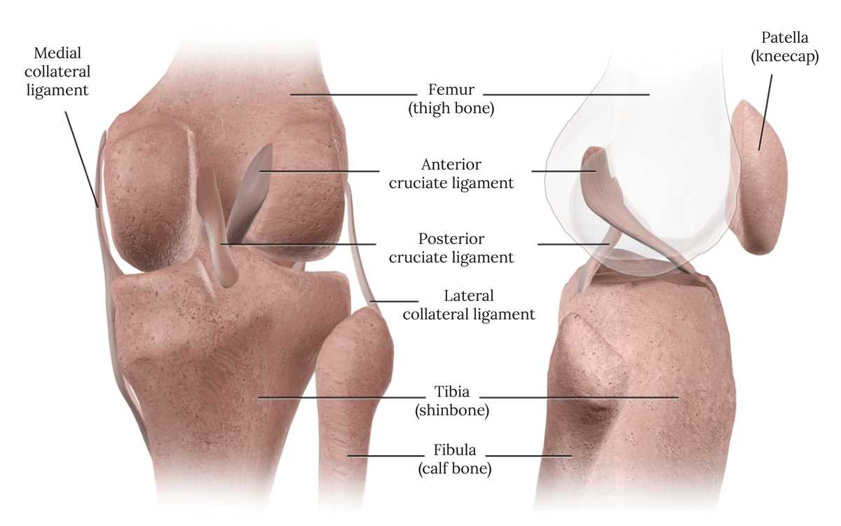
DIS-TANZ DIARY #12

THE KNEE

Mar 15, 2021 in DIS-TANZ-SOLO

Knee Joint Page Title

After taking an in-depth look at shoulder anatomy, movement and strengthening the other day, this time we’ll look at a common weak spot of dancers: the knee.

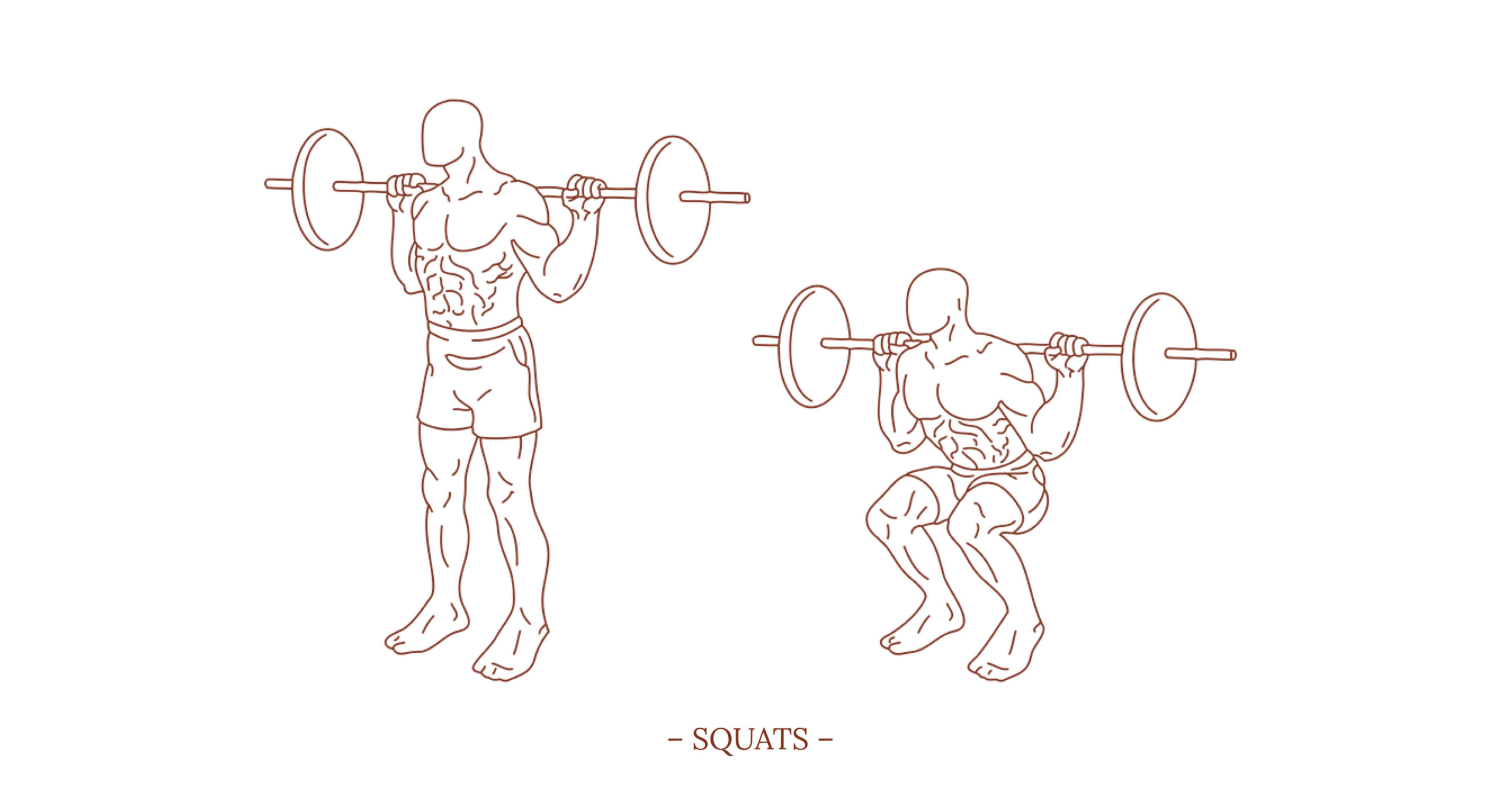
As last time, I would like to recommend the two books ANATOMY OF MOVEMENT by Blandine Calais-Germain and STRENGTH TRAINING ANATOMY by Frédéric Delavier to get even more helpful information on the subject. But let’s dive into the topic…

The Knee

The knee is a complex joint without inherent bony stability. It consists of joints between the femur (thigh bone) and tibia (shinbone) and the patella (kneecap) and femur. It is surrounded by both a fluid-producing layer of synovial tissue and a tough, fibrous capsule.

Ligaments provide support to the connective tissue capsule around the knee. There are four major ligaments that support the knee joint, two collateral ligaments (medial and lateral) and two cruciate ligaments (anterior and posterior). The collateral ligaments are located on both sides of the knee, while the cruciate ligaments cross in the middle of the knee. They form an axis around which the knee rotates.

The Menisci

The two menisci are crescent-shaped discs that act as cushions or "shock absorbers" to allow the bones of the knee to move through their range of motion without rubbing directly against each other. The menisci also contain nerves that help improve balance and stability and ensure proper weight distribution between the femur and tibia. The medial meniscus, is located on the inside of the knee and is the larger of the two. The lateral meniscus lies opposite on the outside of the knee.

Muscles that Cause Movement at the Knee Joint

When fully extended, the tibia and femur "lock" into position, providing stability in the leg and improving weight-bearing capacity. The popliteus muscle, located in the back of the knee, unlocks the knee by rotating the femur on the tibia, allowing the joint to flex.

The hamstring muscle group consists of three muscles on the back of the thigh that affect hip and knee movement. They begin under the gluteus maximus behind the hip bone and attach to the tibia at the knee. The three components are:

1. Biceps femoris – This long muscle flexes the knee. It starts at the thigh and extends to the head of the fibula at the knee.
2. Semimembranosus – This long muscle extends from the pelvis to the tibia. It extends the thigh, flexes the knee, and helps rotate the tibia.
3. Semitendinosus – This muscle also stretches the thigh and flexes the knee, but the tendons that connect it to the bone are much narrower than those of the semimembranosus.

In the calf region of the leg, the gastrocnemius muscle extends from the end of the thigh through the Achilles tendon to the calcaneus. The gastrocnemius forms the posterior muscle wall of the knee and assists in bending the knee.

The quadriceps is a four-headed muscle group on the front of the thigh that does most of the work in extending the knee. These muscles are the strongest and leanest in the entire body. They are:

1. Rectus femoris – In addition to extending the knee, the rectus femoris muscle has additional actions as it is a biarticular muscle that crosses both the hip and knee joints. By acting on the hip joint, it helps flex the thigh. When its insertion is fixed at the patella, this muscle helps flex the pelvis forward toward the thigh. The rectus femoris is also capable of simultaneously flexing the hip and extending the knee.
2. Vastus medialis – This teardrop-shaped muscle of the inner thigh attaches along the thigh and up to the inner edge of the kneecap. It helps with knee extension.
3. Vastus intermedius – Located between the vastus medialis and the vastus lateralis on the front of the thigh, it is the deepest of the four quadriceps muscles.
4. Vastus lateralis – On the outside of the thigh, this is the largest of the four quadriceps muscles. It extends from the top of the thigh at the hip and up to the kneecap.

COMMON INJURIES

MEDIAL KNEE STRAIN

Medial knee strain occurs in the capsule of the medial side of the knee. This injury can often be traced to incorrect positioning of the knee during a plié and typically affects beginners rather than well-trained professional dancers. Accordingly, it usually takes technical training rather than specific medical treatment to fix the problem. In addition, targeted tissue-strengthening training can reduce negative effects that occur due to technique deficiencies.

Patellofemoral Pain Syndrome

Patellofemoral pain syndrome, sometimes called runner's knee, usually causes a dull, aching pain in the front of the knee, especially when walking, climbing stairs, sitting for long periods of time or squatting. Muscle imbalances or weaknesses, as well as overuse, are common causes of this pain in the patellofemoral joint. For most dancers it is enough to lower the load for a while, work on their technique, thereby optimizing the positioning of the kneecap and thus eliminating pain.

Patellar subluxation or dislocation

Patellar subluxation (partial dislocation) or dislocation often occurs due to heavy rotational loading, for example during a twist or turn. As we've already established, several groups of muscles and ligaments hold your kneecap in place. When these are injured, your kneecap can move out of the groove, causing pain and difficulty bending the knee. In most cases, the kneecap is pushed to the outside of the knee, which can correspondingly damage the ligament on the inside. Immediate care should include rest, ice, compression, and elevation (RICE). Further treatment options include nonsteroidal anti-inflammatory medications, physical therapy, crutches or a cane to relieve pressure on the knee, braces or casts to immobilize the knee, and special footwear to relieve pressure on the kneecap. After a first-time patellar subluxation, you have about a 33 percent chance of recurrence.

Patellar tendinitis

Patellar tendinitis is an overuse injury that is also known as jumper's knee. It is an inflammation of the tendon connecting the kneecap to the shinbone. Like all overuse injuries, this injury can be traced back to poor conditioning or wanting too much too fast. Accordingly, the musculoskeletal apparatus is either too weak, not sufficiently warmed up or not flexible enough for the applied load. The stress causes tiny tears in the tendon that your body tries to repair. But as the tears in the tendon multiply, they cause pain due to inflammation and weakening of the tendon. Initial therapy should include cooling with ice and rest to reduce inflammation, further steps include physical therapy to stretch and strengthen the muscles around the knee. Deep squats are recommended as both a post-rehabilitative and preventative measure.

Meniscus Tears

Meniscus tears are among the most common knee injuries among athletes. The medial meniscus is more commonly injured than the lateral meniscus. Any activity where you forcefully twist or rotate your knee, especially if you put full weight on it, can lead to a meniscus tear. But kneeling, squatting low or lifting something heavy are also among the possible causes. Treatment of meniscus tears depends on the size and location of the tear. Other factors that affect treatment include age, activity level and related injuries. The outer portion of the meniscus, often referred to as the "red zone," has good blood supply and can sometimes heal on its own if the tear is small. In contrast, the inner two-thirds of the meniscus, called the "white zone," does not have good blood supply. Tears in this area do not heal on their own because this area lacks blood vessels that supply healing nutrients. If a tear is large and unstable or causes blocking symptoms, surgery may be needed to either repair or remove unstable edges. The procedure is usually fairly simple, and you can often go home the same day. In 85% to 90% of people who have surgery for a meniscal tear, the short-term results are good to excellent. But in the long term, people with a major meniscal injury that is not repairable have a higher risk of developing knee osteoarthritis. Strength training, which focuses on strengthening all structures around the knee joint (muscles, tendons, ligaments), is a good way to take as much load off the meniscus as possible.

Knee ligament injuries

Knee ligament injuries such as strains and tears are fortunately not a very common injury for (contemporary) dancers. Ligament injuries are traumatic injuries that occur when a sudden deforming force exceeds the ligament's ability to lengthen. This can happen, for example, from a sharp change in direction, a false landing from a jump, or most commonly, a blunt force blow to the knee. Such an incident usually has to happen at high speed. The medial collateral ligament and the anterior cruciate ligament are most commonly affected. Treatment of a knee ligament injury depends on the grade of injury, initial steps include the RICE method and administering pain medication. In a mild Grade I sprain, the ligaments may stretch but do not tear. Grade I sprains usually heal within a few weeks. In a moderate Grade II sprain, the knee ligament partially tears. Swelling and bruising are common, and use of the joint is usually painful and difficult. Wearing a weight-bearing brace or supportive taping is a common early treatment. After a Grade II injury, you can usually return to activity once the joint is stable and you are no longer in pain. It can take up to six weeks to heal. In a severe Grade III injury, the ligament tears completely, causing swelling and sometimes bleeding under the skin. As a result, the joint is unstable and cannot bear weight. Often, there is no pain after a Grade III tear because all the pain fibers are torn at the time of injury. Since a grade 3 ligament tear is a complete rupture, the ability to heal itself is much less. Such injuries are long lasting and devastating especially for athletes. Of the 4 knee ligaments, a significant tear of the ACL, PCL, or LCL is more likely to require surgery. However, whether surgical reconstruction of the ligament or attempting rehabilitation by wearing a knee brace is more appropriate is controversial and depends greatly on the individual case.

EXERCISES FOR STRENGTH AND STABILITY

The most important weight-bearing joints in the body are the knees, ankles and hips. When you walk across a flat surface, the force on your knees is already about 1.5 times your body weight. For a simple vertical jump, the load increases to 6.9 to 9.0 times your body weight (Cleather, Goodwin, and Bull, 2013 – earlier studies in this field suggested values as high as 20 times the body weight). Accordingly, strong, balanced muscles can help take a lot of strain off your knees.

To prevent overuse damage and to protect and strengthen your knees in general, you should focus on movements that work the hamstrings, quadriceps, glutes, hip muscles, and gastrocnemius.

To be clear, the following exercises are primarily aimed at healthy dancers who want to work preventively. Those who already have knee problems and are looking for a way to get their bodies back in shape should probably start with lighter exercises. I definitely recommend contacting a health or sports science expert to analyze the specific case and set up an appropriate training plan.

1. SQUATS
– targets mainly the quadriceps, gluteal muscles, adductor group, erector spinae, abdominal muscles, and the hamstrings

2. DUMBBELL SQUATS
– targets mainly the quadriceps and the gluteal muscles

3. INCLINE LEG PRESSES
– targets mainly the quadriceps, gluteal muscles, and adductors (depending on the positioning of the feet)

4. BARBELL LUNGES / DUMBBELL LUNGES
– targets mainly the gluteus maximus, quadriceps and the hamstrings

5. GOOD MORNINGS
– targets mainly the hamstrings, gluteus maximus, and the spinal group

6. CABLE BACK KICKS / MACHINE HIP EXTENSIONS
– targets mainly the gluteus maximus, and to a lesser extent the hamstrings

7. BRIDGING
– targets mainly the gluteus maximus and the hamstrings

8. LEG EXTENSIONS
– targets mainly the quadriceps

9. LYING LEG CURLS
– targets mainly the hamstrings, gastrocnemius, and popliteus

10. STANDING LEG CURLS
– targets mainly the hamstrings and to a lesser extent the gastrocnemius

11. SEATED LEG CURLS
– targets mainly the hamstrings, popliteus, and to a lesser extent the gastrocnemius

12. CABLE HIP ADDUCTION / MACHINE HIP ADDUCTION
– targets mainly the adductor group

13. CABLE HIP ABDUCTION / MACHINE HIP ABDUCTION
– targets mainly the gluteal muscles

14. STANDING CALF RAISES
– targets mainly the triceps surae (i.e. soleus and gastrocnemius)

15. SEATED MACHINE CALF RAISES / SEATED BARBELL CALF RAISES
– targets mainly the soleus

FURTHER READING

Acta Orthopedica Cover

Anterior cruciate ligament injury in professional dancers

by Duncan E. Meuffels & Jan A. N. Verhaar
(Acta Orthopaedica 79/4, 2008)

Anterior cruciate ligament injury (ACL) is a common sport injury; however, there are no data concerning dance and ACL injury. We report the incidence, injury mechanism, and clinical follow-up of ACL injury in professional dancers. In a retrospective cohort study involving the three major dance companies in the Netherlands, by interviewing all 253 dancers who had had a full-time contract during 1991-2002, dancers with symptomatic ACL injury or past ACL reconstruction were identified and examined.

British Journal of Sports Medicine Cover

Gluteal muscle activity and patellofemoral pain syndrome: a systematic review

by Christian J Barton, Simon Lack, Peter Malliaras & Dylan Morrissey
(British Journal of Sports Medicine 47/4, March 2013)

There is growing evidence to support the association of gluteal muscle strength deficits in individuals with patellofemoral pain syndrome (PFPS) and the effectiveness of gluteal strengthening when treating PFPS. In addition, an impressive body of work evaluating gluteal electromyography (EMG) has recently emerged, further supporting the importance of gluteal muscle function in PFPS. This systematic review synthesises these EMG findings in order to better understand the role of gluteal muscle activity in the aetiology, presentation and management of PFPS.

Clinical Biomechanics Cover

Hip and knee joint loading during vertical jumping and push jerking

by Daniel J. Cleather, Jon E. Goodwin & Anthony M. J. Bull
(Clinical Biomechanics 28/1, January 2013)

The internal joint contact forces experienced at the lower limb have been frequently studied in activities of daily living and rehabilitation activities. In contrast, the forces experienced during more dynamic activities are not well understood, and those studies that do exist suggest very high degrees of joint loading. Methods: In this study a biomechanical model of the right lower limb was used to calculate the internal joint forces experienced by the lower limb during vertical jumping, landing and push jerking (an explosive exercise derived from the sport of Olympic weightlifting), with a particular emphasis on the forces experienced by the knee.

ournal of Orthopaedic & Sports Physical Therapy Cover

Isokinetic Characteristics of the Knee in Female, Professional and University, Ballet and Modern Dancers

by Robin D. Chmelar, Barry B. Shultz, Robert R. Ruhling, Sally S. Fitt & Mary B. Johnson
(Journal of Orthopaedic & Sports Physical Therapy 9/12, June 1988)

The purpose of this investigation was to evaluate isokinetic characteristics of the knee in female, ballet and modern, professional and university dancers in order to evaluate possible differences among the groups. A total of 37 dancers with a mean age of 24.9 years was tested using a Cybex II® dynamometer. A multiple analysis of variance (MANOVA) indicated that the ballet dancers had significantly higher H/Q ratios than modern dancers at three speeds (p < 0.024). Post hoc procedures indicated that the professional ballet dancers (PB) had significantly higher H/Q ratios than all other groups at 180°/sec (p < 0.05). Also, although most of these dancers demonstrated normal peak torque/body weight values for knee extension and flexion, specific weaknesses were observed in the force decay rate of the quadriceps curves. It was concluded that these theatrical dancers were not a homogeneous group in terms of certain isokinetic characteristics.

Prevention of Injuries in the Young Dancer Cover

Knee Problems in the Young Dancer

by Peter G. Gerbino, Andrea Stracciolini & Marina G. Gearhart
(Prevention of Injuries in the Young Dancer, 2017)

Young dancers are at unique risk for knee injuries as the result of the inherent demands of dance and the biomechanical changes that occur during growth. The majority of knee injuries in young dancers are due to overuse, as opposed to macrotrauma. Since many young dancers have joint hypermobility and disordered eating patterns, these variables play a large role in the injury profile for this population. Bone health, including proper nutrition and vitamin D levels, is another unusually important variable to be considered in the assessment of dance injuries. This chapter deals with many of the knee injuries that young dancers commonly sustain, such as patellofemoral pain syndrome, fat pad syndrome, plica syndrome, Osgood–Schlatter syndrome, and Sinding–Larsen–Johansson syndrome. As with all activity-related injuries, the first task in dealing with these injuries is to make an accurate diagnosis. Next, the pathophysiology needs to be understood, as well as the risk factors involved. Treatment, and finally prevention, completes the medical management for optimal care.

NAJSPT Article Preview

Rehabilitation of a Female Dancer with PatellOfemoral Pain Syndrome: Applying Concepts of Regional Interdependence in Practice

by Caitlyn Welsh, William J. Hanney, Laura Podschun & Morey J. Kolber
(North American Journal of Sports Physical Therapy 5/2, June 2010)

Due to complex movements and high physical demands, dance is often associated with a multitude of impairments including pain of the low back, pelvis, leg, knee, and foot. This case report provides an exercise progression, emphasizing enhancement of strength and neuromuscular performance using the concept of regional interdependence in a 17 year old female dancer with patellofemoral pain syndrome.

Header photo (Muscles, tendons and bones of the knee-joint: two figures. Red chalk and pencil drawing by or associated with A. Durelli, ca. 1837) courtesy of Wellcome Collection, Attribution 4.0 International (CC BY 4.0), Edited by Michael Loehr

Anatomy of the knee illustrations courtesy of Blausen.com staff / Medical gallery of Blausen Medical 2014 / WikiJournal of Medicine, CC BY 3.0, via Wikimedia Commons, Edited by Michael Loehr

Exercises for strength and stability illustrations courtesy of Everkinetic, CC BY-SA 3.0, via Wikimedia Commons, Edited by Michael Loehr

DIS-TANZ-SOLO

Overview

DIS-TANZ-SOLO Research Bibliography

Bibliography

DIS-TANZ-SOLO Research Links

Links

Gefördert durch die Beauftragte der Bundesregierung für Kultur und Medien im Programm NEUSTART KULTUR, Hilfsprogramm DIS-TANZEN des Dachverband Tanz Deutschland.

Logo Die Beauftragte der Bundesregierung für Kultur und Medien
Logo Neustart Kultur
Logo Dachverband Tanz Deutschland
Logo Dis-Tanzen