DIS-TANZ DIARY #8

THE SHOULDER

Feb 15, 2021 in DIS-TANZ-SOLO

Shoulder joint Page Preview Photo

As I said from the beginning, this diary will be quite subjective and erratic. I’m taking you along on my personal research and learning journey, sharing all the information that seems interesting as it comes.

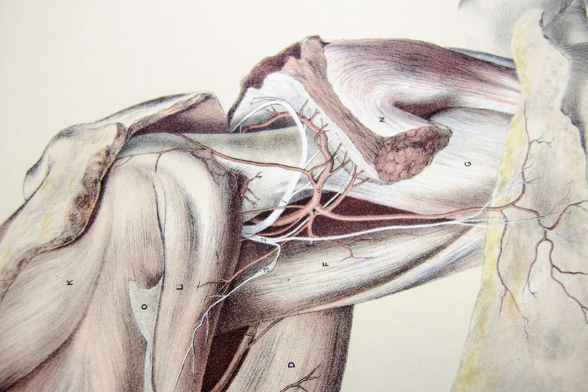
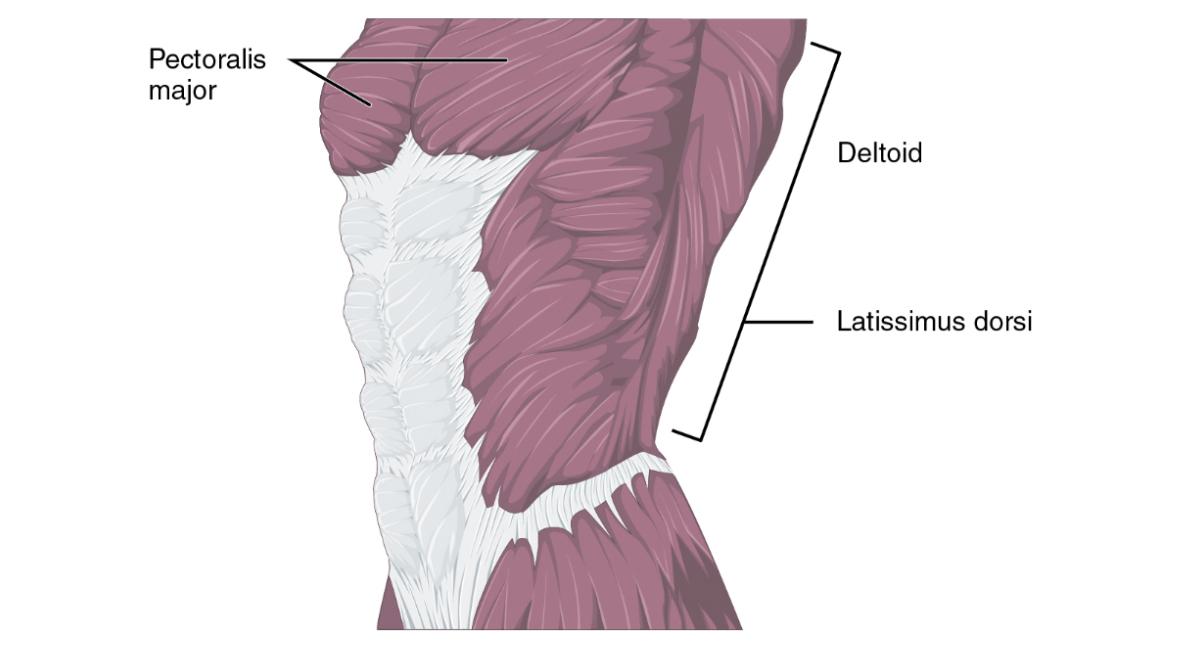
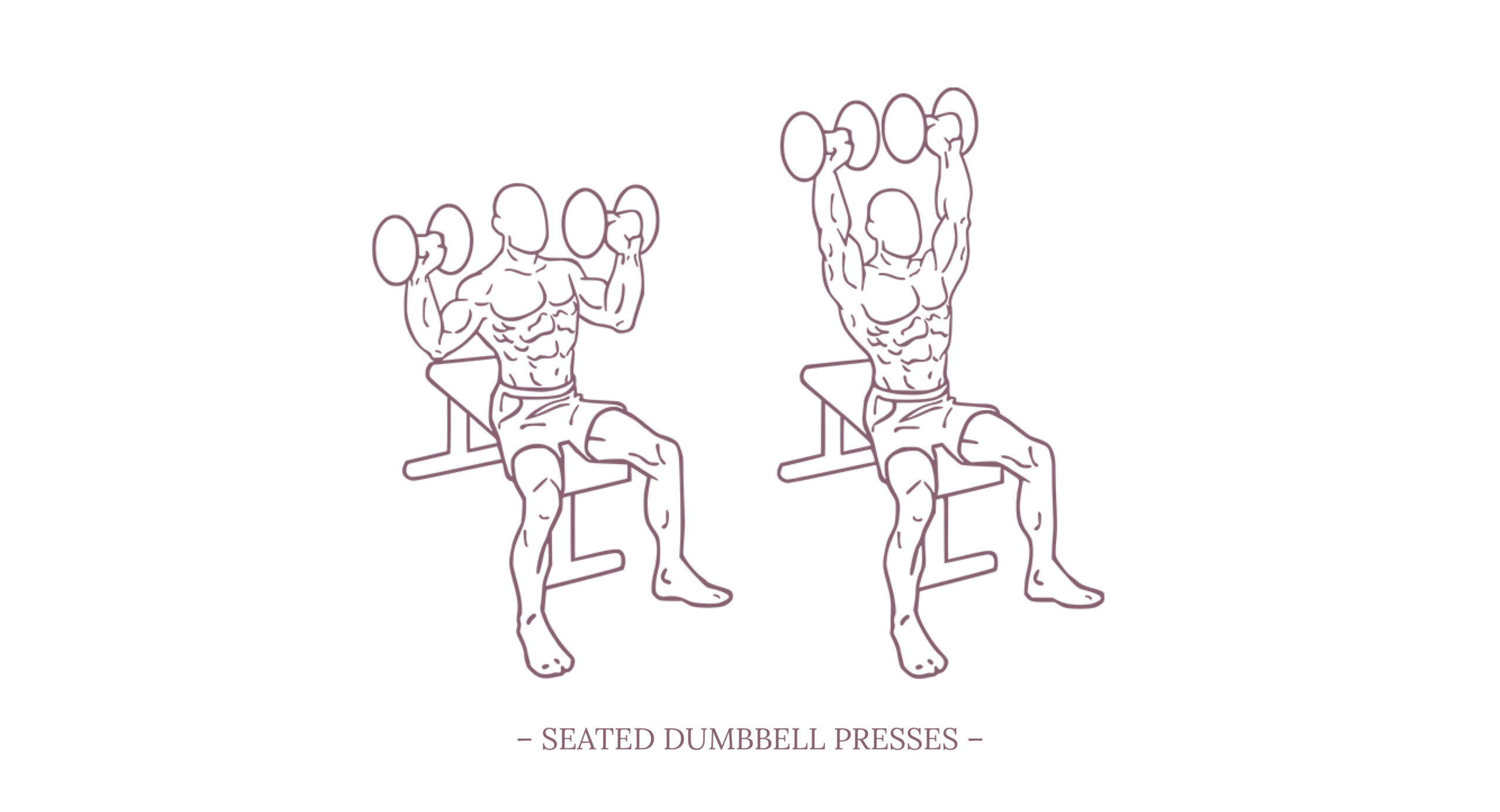
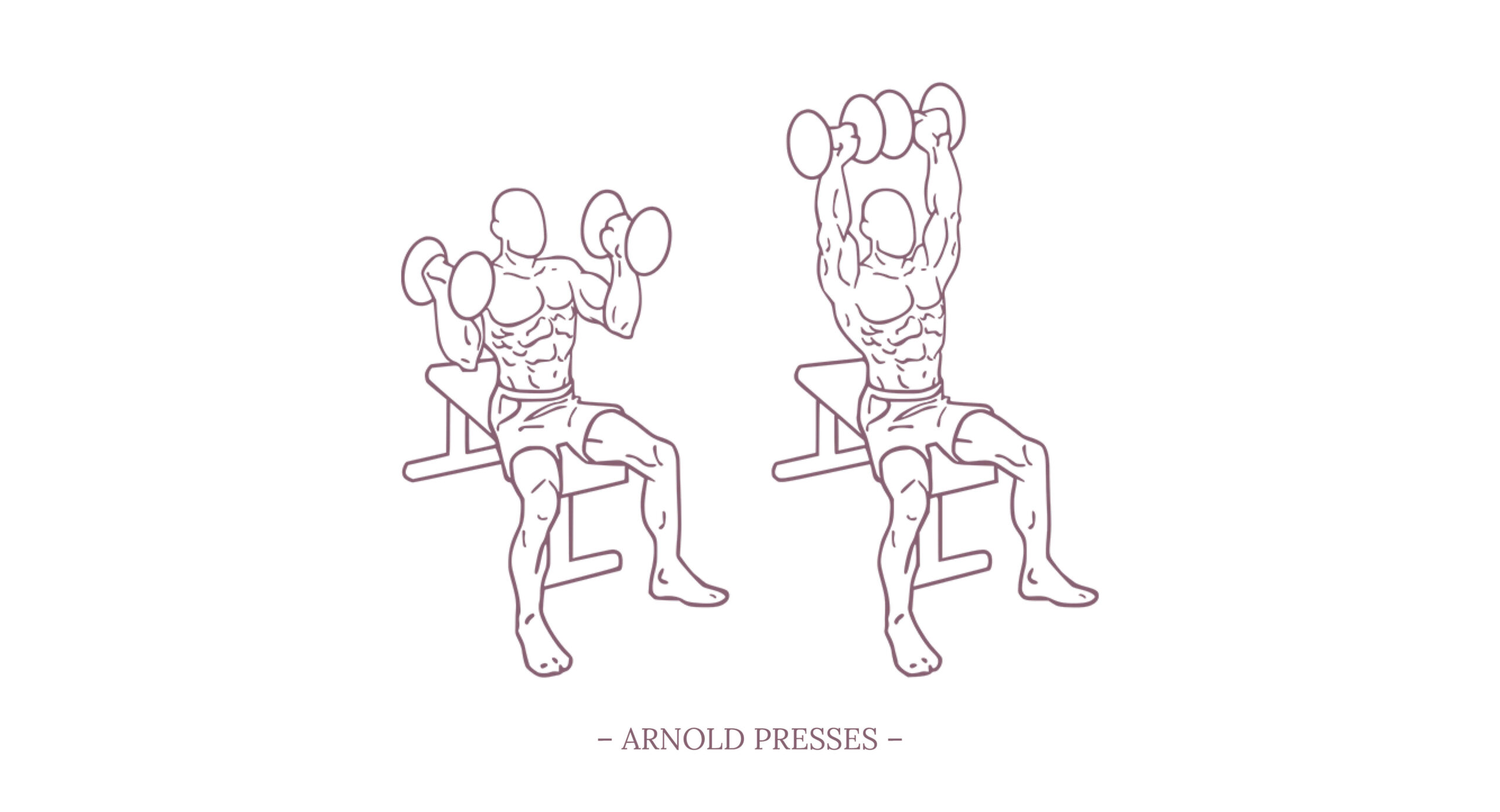
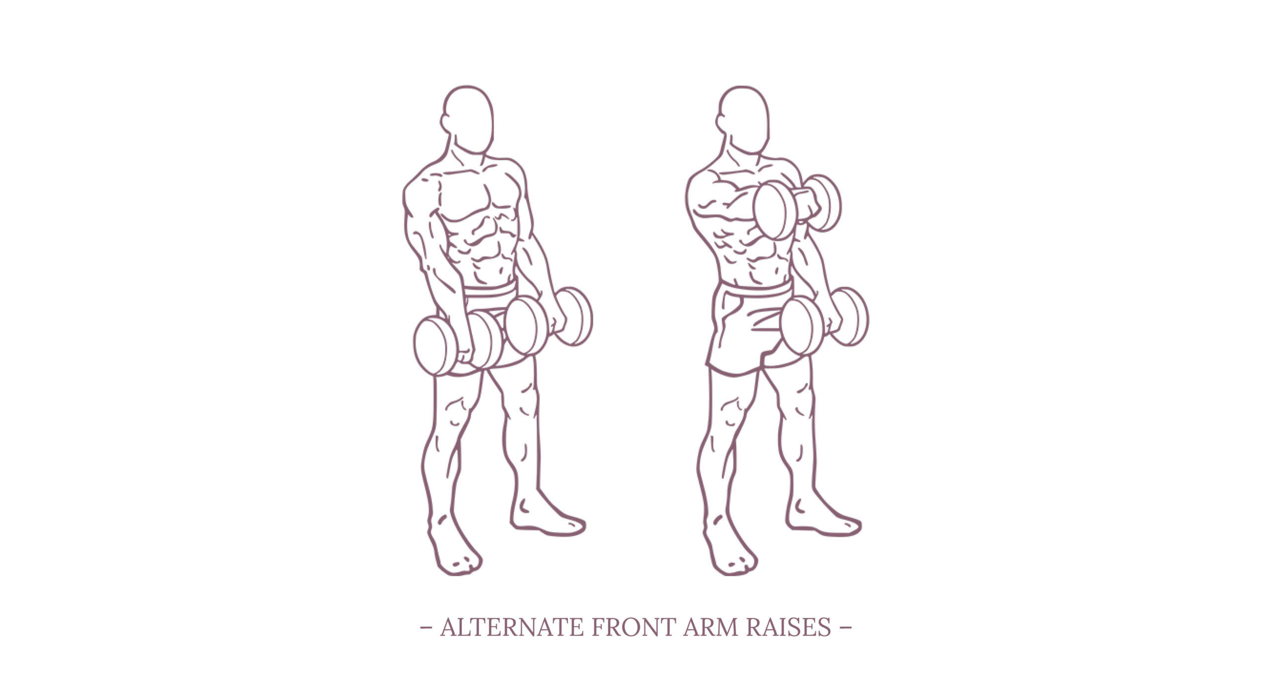
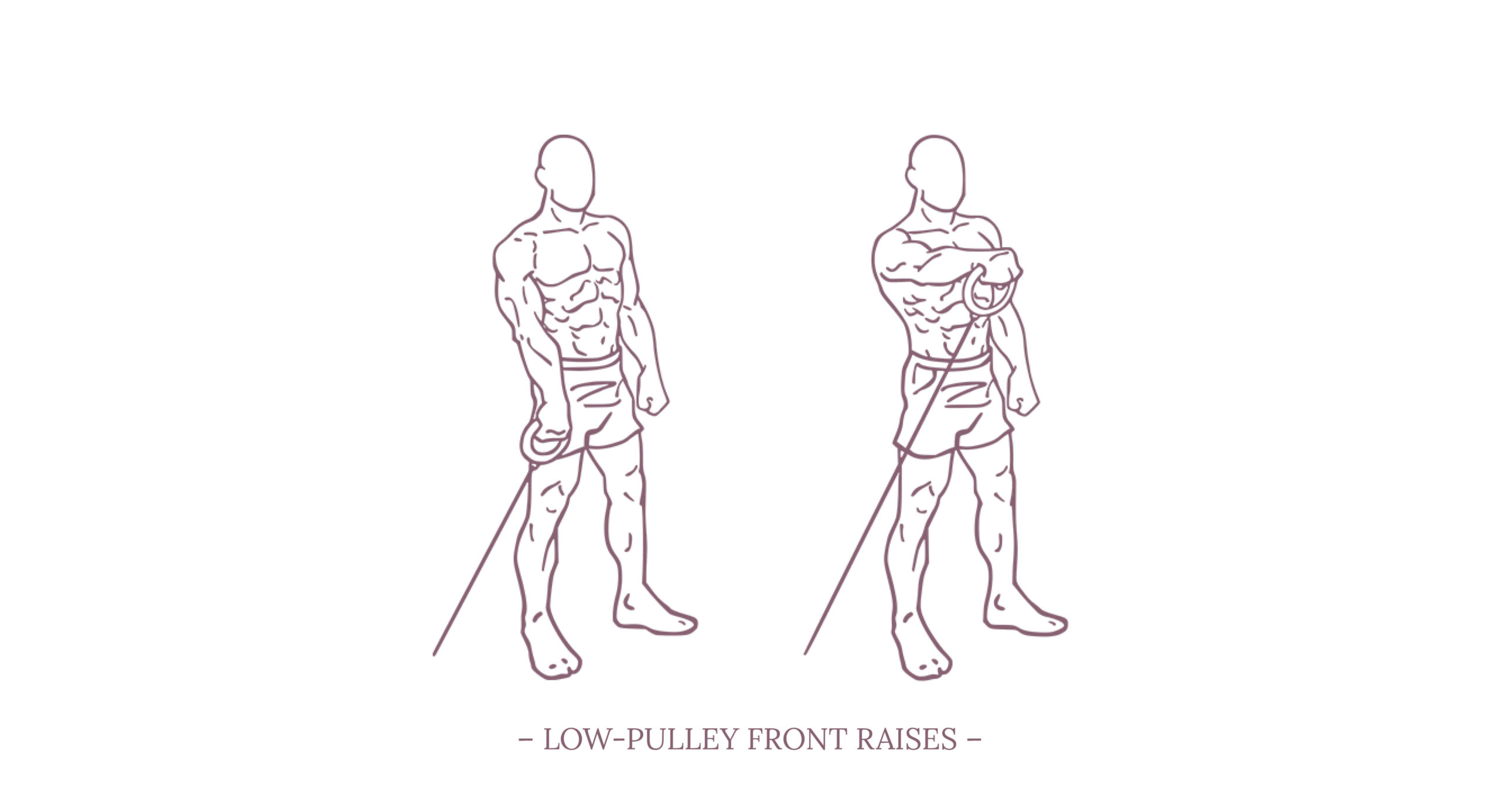
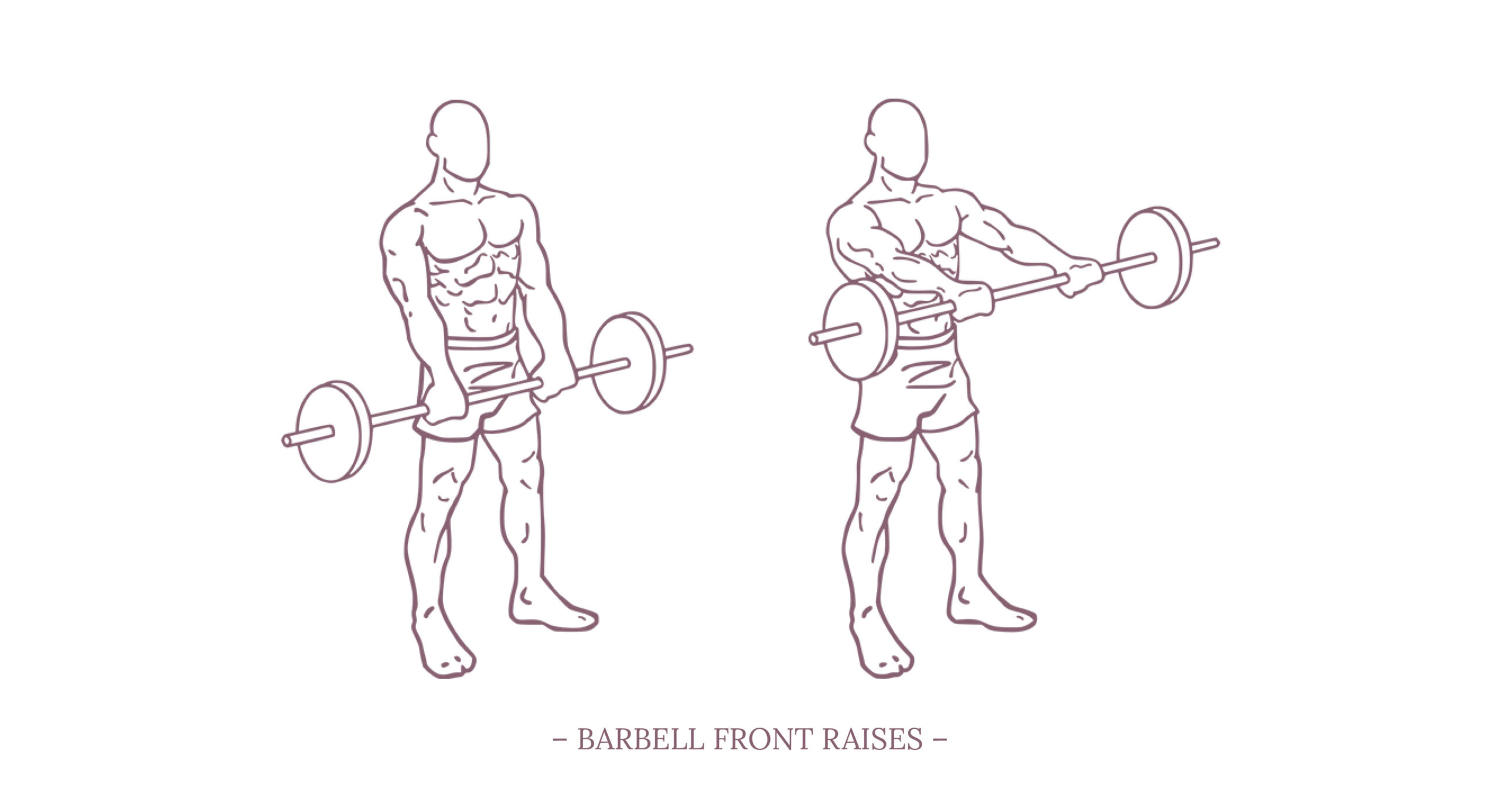
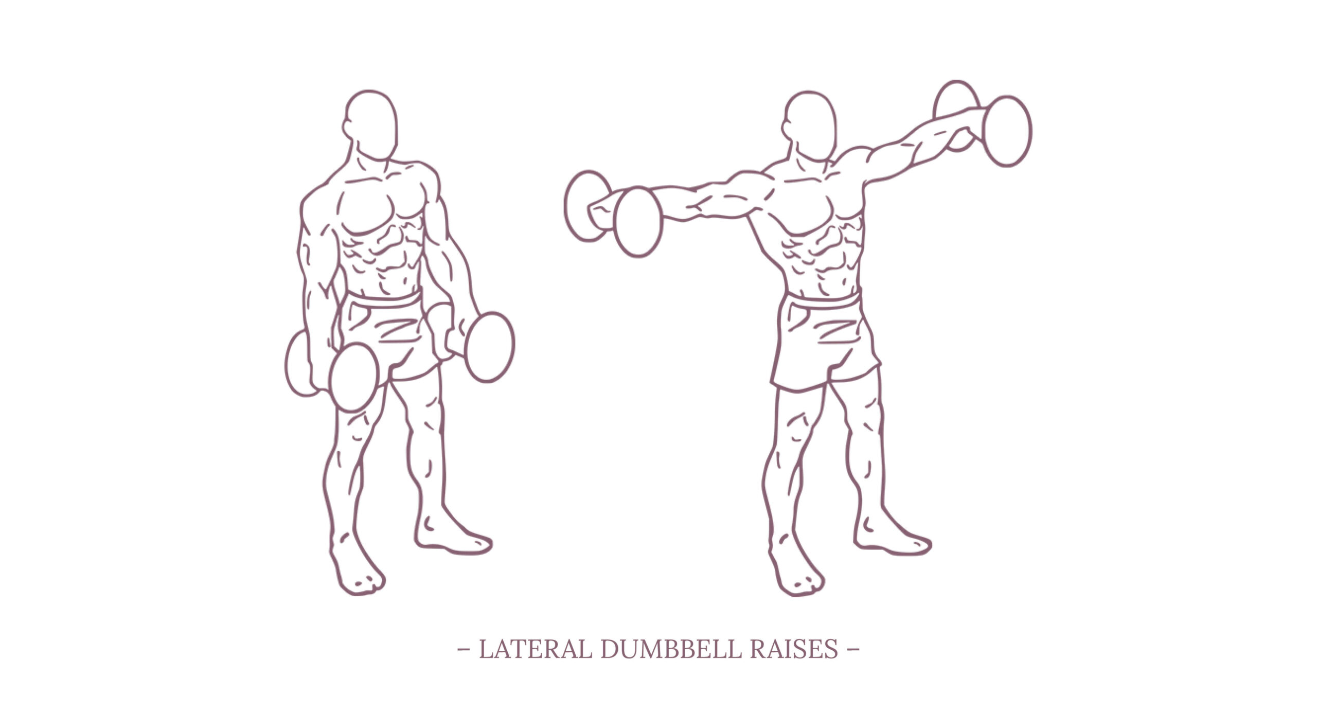
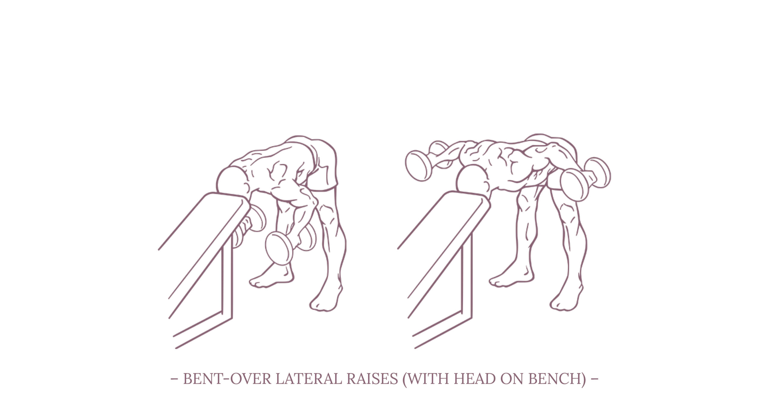
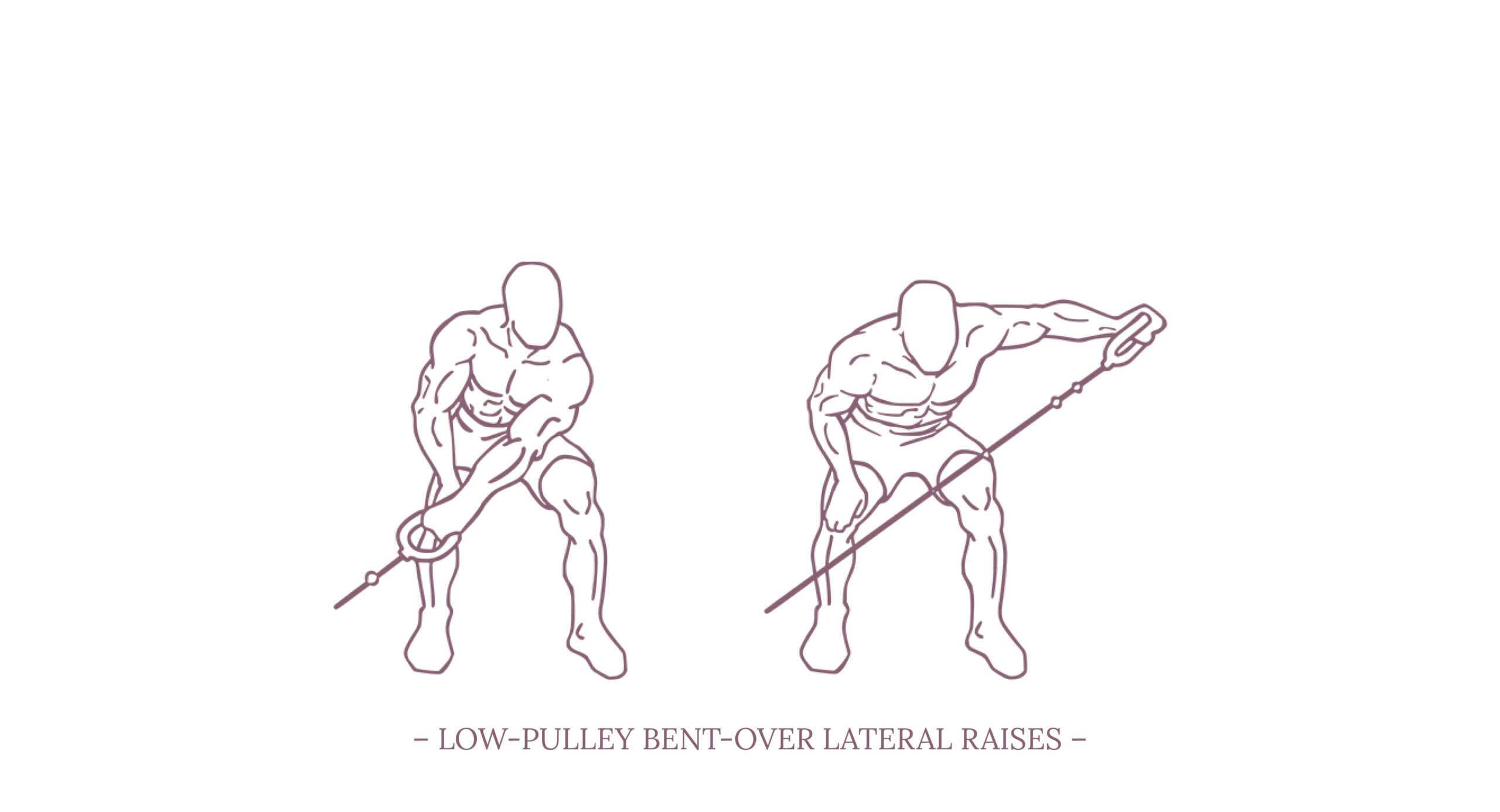
Since I’m currently dealing with the last fades of a nagging shoulder problem, and therefore dealing with anatomy, physiology, and appropriate strengthening exercises, this chapter is a bit of a digression on all things shoulder.

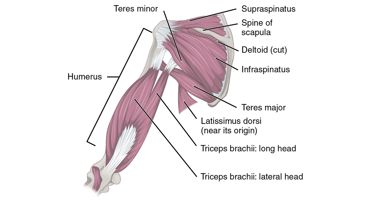
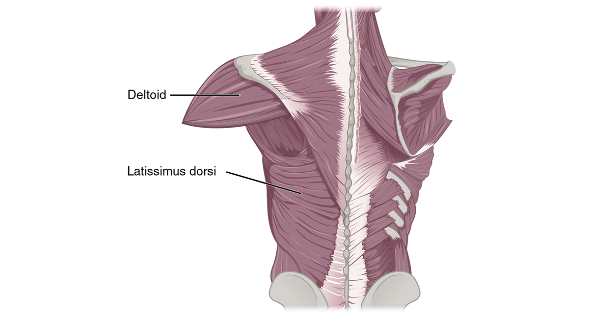
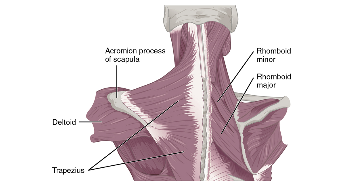
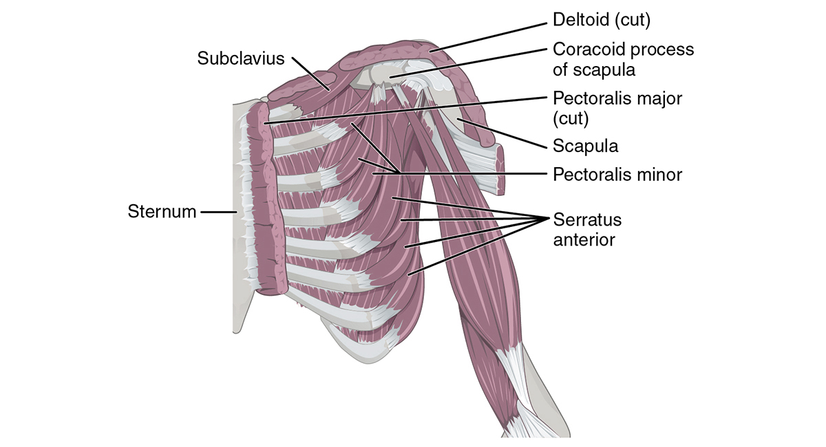
I will focus mainly on the anatomical principles that are directly relevant to our work, i.e. bone and muscle structures, and won’t touch on neurovascular aspects.

If you are looking for more in-depth information on the subject, I highly recommend ANATOMY OF MOVEMENT by Blandine Calais-Germain and STRENGTH TRAINING ANATOMY by Frédéric Delavier. Both books combined give a comprehensive impression of anatomy as well as practical applications that are super useful for our work in the dance studio as well as in the gym.

The Shoulder

The shoulder is one of the largest and most complex joints in the body. When you get right down to it, the shoulder consists not of just one joint, but of various joints that work together in a complex way to enable the arm's wide range of motion.

The shoulder is formed by different bones: the upper arm bone (humerus), the shoulder blade (scapula) and the collarbone (clavicle). These bones also form the various joints of the shoulder:

1. The glenohumeral joint – the joint between the humeral head and the glenoid cavity of the scapula
2. The acromioclavicular joint – the joint formed by the acromion and the clavicle
3. The sternoclavicular joint – the joint between the clavicle and the sternum
4. The scapulothoracic joint – the articulation between the scapula and the back of the rib cage (thorax), not a true synovial joint

The Glenohumeral Joint

The glenohumeral joint, sometimes simply referred to as the shoulder joint, is the main joint of the shoulder and its anatomy is quite unique - structurally it is a ball and socket joint between the scapula and the humerus. Due to the relative size of the humeral head compared to the shallow glenoid cavity and its loose joint capsule, it allows flexibility and range of motion not found elsewhere in the body.

However, this exceptional shoulder function comes at the expense of stability. Because it lacks strong ligaments and its bony structures offer little support, the shoulder joint is known as a muscle-dependent joint. It is mainly stabilized by the muscles and ligamentous structures of the so-called rotator cuff.

The Rotator Cuff

The rotator cuff consists of four muscles: subscapularis, supraspinatus, infraspinatus, and teres minor. Each of these four muscles originates at the scapula and has a tendon that attaches to the head of the humerus.

As a group, the rotator cuff muscles are responsible for stabilizing the shoulder joint by providing fine-tuning of the movements of the humeral head within the glenoid cavity. They are deeper muscles and very actively involved in the neuromuscular control of the shoulder during upper extremity movements.

Cranial to the rotator cuff (i.e., toward the head end of the body) is a bursa that covers and protects the muscle and tendons as they are in close contact with the surrounding bones.

The supraspinatus primarily abducts the shoulder and is responsible for the initial 15 degrees of abduction. The infraspinatus and teres minor aid in external rotation of the shoulder. The subscapularis muscle aids in internal rotation of the shoulder.

The "Global Movers"

In addition to the aforementioned fine stabilizers, there are a number of other muscles that can be categorized as the global movers of the shoulder. The larger muscles such as the trapezius, levator scapula, pectorali, deltoids, serratus anterior, latissimus dorsi, rhomboids, teres major, biceps, coracobrachialis, and triceps muscles work together as agonist and antagonist couplings to enable the gross motor movements of the entire shoulder region.

Movements of the shoulder

As described above, the glenohumeral joint has the ability to allow an extreme range of motion in multiple directions.

Flexion is defined as bringing the upper limb forwards in the sagittal plane (i.e., moving the arm forwards). The usual range of motion is 180 degrees. The main flexors of the shoulder are the anterior deltoid, coracobrachialis, and pectoralis major. The Biceps brachii weakly assists in this action.

Extension is defined as bringing the upper limb backwards in a sagittal plane (i.e.; moving the arm backwards). The normal range of motion is 45 to 60 degrees. The main extensors of the shoulder are the posterior deltoid, latissimus dorsi, and teres major.

Abduction is defined as bringing the upper limb away from the midline in the coronal plane (i.e., moving the arm sideways, away from the body). The normal range of motion is 150 degrees. The first 0-15 degrees of abduction is produced by the supraspinatus. The middle fibers of the deltoid are responsible for approximately 15 to 90 degrees of abduction following. Past 90 degrees, the scapula needs to be rotated to achieve abduction; this is carried out by the trapezius and serratus anterior.

Adduction is defined as bringing the upper limb towards the midline in the coronal plane (i.e., moving the arm sideways, towards the body). Pectoralis major, latissimus dorsi, and teres major are the muscles primarily responsible for shoulder adduction.

Internal rotation is defined as rotation toward the midline along a vertical axis, so that the thumb is pointing inwards. The normal range of motion is 70 to 90 degrees. The internal rotation muscles are the subscapularis, pectoralis major, latissimus dorsi, teres major, and the anterior aspect of the deltoid.

External rotation is defined as rotation away from the midline along a vertical axis, so that the thumb is pointing outwards. The normal range of motion is 90 degrees. Primarily infraspinatus and teres minor are responsible for the motion.

Impingement Syndrome

The fact that shoulders are relatively low on the injury scale for dancers is due less to good training than to the fact that they are subjected to relatively little stress.

One of the more common injuries is the shoulder impingement syndrome; it is usually characterized by pain during overhead movements or when lifting objects. The shoulder impingement syndrome is primarily caused by a narrowing of the space below a bony process (the acromion) in the shoulder. The supraspinatus muscle (one of the rotator cuff muscles) runs through this space and can become inflamed when compressed. In some cases, the supraspinatus bursa also becomes compressed and inflamed, or calcium deposits may form within the supraspinatus tendon.

Triggers may include frequently repeated arm movements, especially in overhead activities, such as frequent extension of the arm at high speed under high loads or repeated lifts. Increased upper extremity training by a dancer whose rotator cuff muscles are not in good condition can also be a reason for shoulder impingement.

Initial treatment for shoulder impingement includes ice, rest and taking anti-inflammatory medications to reduce acute swelling in the joint. For chronic cases, the doctor may recommend a corticosteroid injection to reduce inflammation in the joint. Physical therapy or athletic consultation is recommended to assess shoulder function and improve rotator cuff strength. Dancers with chronic, unresolved impingement symptoms may be candidates for a surgical technique to decompress the acromioclavicular joint.

Shoulder exercises for Strength and Stability

Here is a selection of classic exercises that strengthen and stabilize the shoulder. These exercises are designed for the use of dumbbells, barbells, or pulley systems, but most of these can be adapted for a training with resistance bands as well.

1. SEATED FRONT PRESSES
– targets mainly the anterior and lateral deltoids, the clavicular head of the pectoralis major, triceps bracchi, serratus anterior, trapezius, and supraspinatus

2. SEATED DUMBBELL PRESSES
– targets mainly the middle deltoid, trapezius, serratus anterior, and triceps bracchi

3. ARNOLD PRESSES*
– targets mainly the anterior deltoid, the clavicular head of the pectoralis major, triceps bracchi, trapezius, and serratus anterior

4. ALTERNATE FRONT ARM RAISES / LOW-PULLEY FRONT RAISES
– targets mainly the anterior deltoid, and the clavicular head of the pectoralis major

5. BARBELL FRONT RAISES
– targets mainly the anterior deltoid, the clavicular head of the pectoralis major, and infraspinatus

6. ONE-DUMBBELL FRONT RAISES
– targets mainly the anterior deltoid, the clavicular head of the pectoralis major, and the short head of the biceps

7. LATERAL DUMBBELL RAISES / LOW-PULLEY LATERAL RAISES
– targets mainly the middle deltoid

8. MACHINE LATERAL RAISES
– targets mainly the deltoid and supraspinatus

9. BENT-OVER LATERAL RAISES / LYING LATERAL RAISES
– targets mainly the posterior deltoid, trapezius, rhomboids, teres minor, and infraspinatus

10. LOW-PULLEY BENT-OVER LATERAL RAISES
– targets mainly the posterior deltoids, trapezius, and the rhomboids

11. HIGH-PULLEY LATERAL EXTENSIONS
– targets mainly the posterior deltoid, infraspinatus, teres minor, trapezius, and the rhomboids

12. PEC DECK REAR-DELT LATERALS (REVERSE FLYS)
– targets mainly the posterior deltoid, infraspinatus, teres minor, trapezius, and rhomboids

13. FACE PULLS
– targets mainly the posterior deltoid, trapezius, rhomboids, infraspinatus, and teres minor

14. EXTERNAL ARM ROTATIONS AT A PULLEY
– targets mainly the infraspinatus

* The Arnold Press is an incredibly effective shoulder exercise that can provide a very strong stimulus for generating functional strength and hypertrophy in the deltoids and arms. However, there are common mistakes that many lifters make that not only shorten the benefits of this exercise, but can also lead to joint problems and inflammation. So the exercise should only be performed as an advanced lifter, and is less suitable as recovery training after impingement.

Header photo courtesy of University of Liverpool Faculty of Health & Life Sciences from Liverpool, United Kingdom, CC BY-SA 2.0, via Wikimedia Commons

Muscles that position the pectoral girdle & Muscles that move the humerus illustrations courtesy of OpenStax College: Anatomy & Physiology, CC BY 3.0, via Wikimedia Commons (Edited by Michael Loehr)

Shoulder strength exercise illustrations courtesy of Everkinetic, CC BY-SA 3.0, via Wikimedia Commons (Edited by Michael Loehr)

DIS-TANZ-SOLO

Overview

DIS-TANZ-SOLO Research Bibliography

Bibliography

DIS-TANZ-SOLO Research Links

Links

Gefördert durch die Beauftragte der Bundesregierung für Kultur und Medien im Programm NEUSTART KULTUR, Hilfsprogramm DIS-TANZEN des Dachverband Tanz Deutschland.

Logo Die Beauftragte der Bundesregierung für Kultur und Medien
Logo Neustart Kultur
Logo Dachverband Tanz Deutschland
Logo Dis-Tanzen